Histology, Imaging and Single Cell Sequencing
Tissue, Imaging & Single-Cell Services
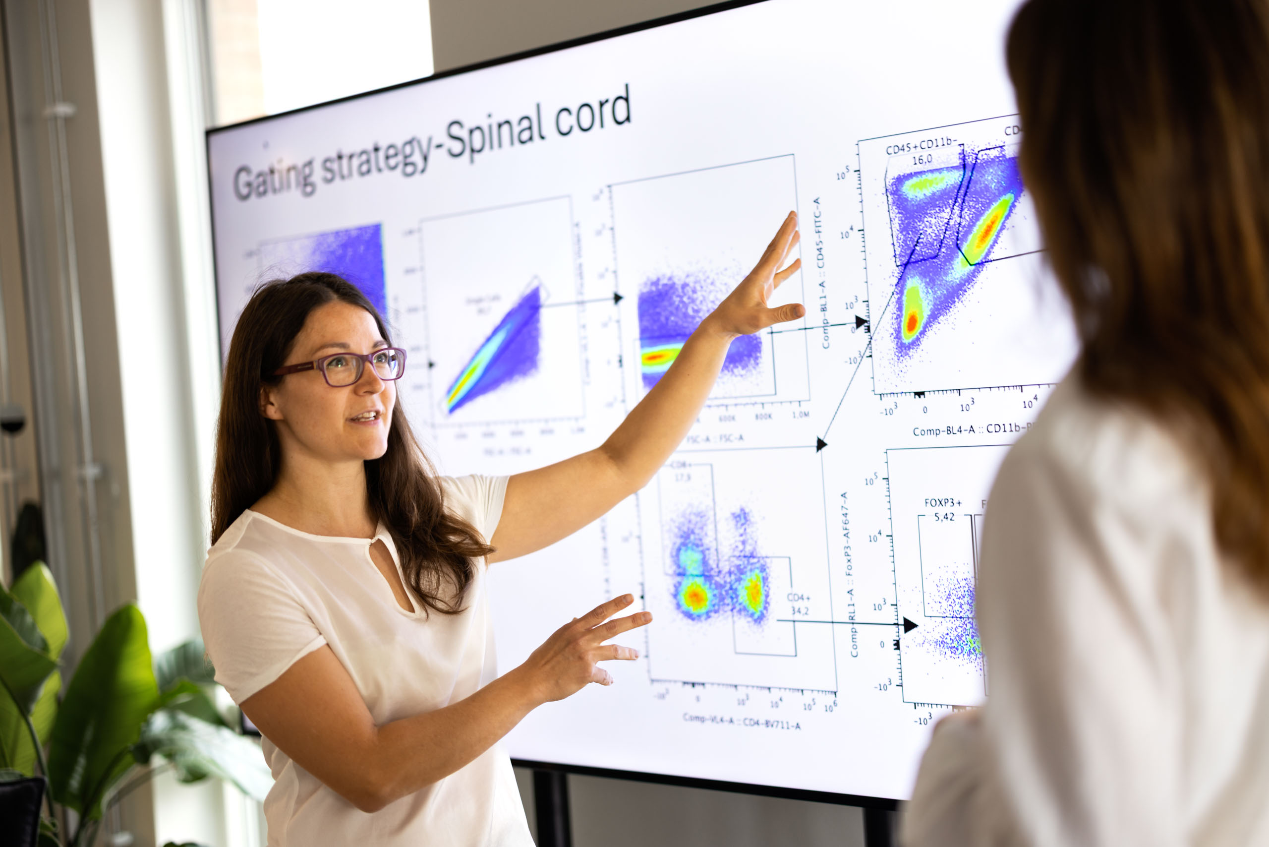
Understanding how a therapy modulates disease requires more than functional readouts — it demands spatial, cellular, and molecular resolution. Tissue analysis, advanced imaging, and single-cell sequencing provide critical insight into mechanism, target engagement, and cellular dynamics within complex biological systems.
At Redoxis, we integrate quantitative histopathology, high-resolution imaging, and single-cell transcriptomics to generate multidimensional data with strong translational relevance. From tissue architecture and immune cell infiltration to cell-specific gene expression signatures, our analyses deepen mechanistic understanding and strengthen preclinical decision-making.
- Quantitative histopathology and blinded scoring
- H&E and special stainings
- Immunohistochemistry (IHC) and immunofluorescence (IF)
- Multiplex tissue staining
- Digital slide scanning and image analysis
- Single-cell RNA sequencing (scRNA-seq)
- Immune cell subset identification and transcriptomic profiling
- Bioinformatic analysis and pathway enrichment
- In vivo analysis of behaviour and gait
Seamlessly connected to our in vivo and in vitro platforms, these services complete the biological picture — delivering robust, decision-enabling insight to advance your development program with confidence.
Contact us
Nina Woodworth
COO